Showing posts with label orthopedic doctor apollo hospital. Show all posts

Tuesday, July 11, 2023

How to Identify Health Issues of Vitamin D Deficiencies ?

 Vitamin D is essential for strong bones. It helps the body absorb calcium from the food that we eat. Lack of vitamin D can lead to bone diseases such as rickets and osteoporosis. But did you know that deficiency of vitamin D can also affect your overall health and well-being?

Dr. A. Mohan Krishna, an orthopedic surgeon at Apollo Hospitals, suggests that being aware of the following symptoms can help identify vitamin D deficiencies:

1. Muscle Weakness: Vitamin D is necessary for the proper functioning of muscles. Deficiency of vitamin D can lead to muscle weakness and aches in various parts of the body.

2. Chronic Pain: Muscle and joint pain, particularly in the lower back and knees, can be a sign of vitamin D deficiency.

3. Mood Swings: Vitamin D plays a crucial role in the production of serotonin, a hormone responsible for regulating mood. Low levels of vitamin D can lead to mood swings and depression.

4. Frequent Infections: Vitamin D is vital for a strong immune system. A deficiency can make you susceptible to infections like cold and flu.

If you experience any of the above symptoms, it's essential to get tested for vitamin D deficiency. This can be done through a simple blood test. If diagnosed with a deficiency, your doctor may recommend taking supplements or increasing sun exposure.

Don't ignore the signs of vitamin D deficiency. Take care of your bones and overall heal

Wednesday, April 13, 2016

CARPAL TUNNEL SYNDROME


Carpal tunnel syndrome:

The carpal tunnel is the space between the bones and ligaments of the wrist through which median nerve passes. Carpal tunnel syndrome develops when the nerve becomes compressed. It is common condition of the hand that affects women more frequently than men.

The symptoms of carpal tunnel syndrome include:
-        
  Pins and needles or tingling sensations (Paraesthesias)
-         
Pain or numbness in the hand, typically worse at the night.
-         
Clumsiness and weakness of the hand.
-         
A weak grip and an impaired ability to bring the thumb across the palm to meet the other fingers.
-         
Pain in the wrist, forearm or shoulder.

Causes:

Any condition that narrows the carpal tunnel or produces swelling of or fluid retention by the contents of the tunnel can cause carpal tunnel syndrome. The many possible causes include

-          Hormonal changes.
-          Obesity,
-          Diabetes mellitus,
-          Rheumatoid arthritis,
-          Acromegaly – bone enlargement due to pituitary gland abnormality.
-          Under activity of thyroid (hypothyroidism)
-          Renal failure,
-          Alcoholism
-          Amyloidosis : rare condition in which abnormal proteins accumulate in tissues and organs.
-          Paget’s disease: a chronic bone disease that affects elderly people. The bones become deformed and thickened.
-          Tumors: such as lipoma (Fatty tumours), ganglions (fluid filled cysts formed in tendon), and deformities of wrist after the fractures.
-          The use of hand held vibrating tools – very rarely causes carpal tunnel syndrome.

Diagnosis:

The typical history of pain and weakness in the hands usually suggest the diagnosis of carpal tunnel syndrome, but it is important to exclude other conditions that may produce similar symptoms, such as a prolapsed cervical disc or arthritis of thumb joint.

Clinical examination:

This may reveal disturbances in sensation in the area supplied by the median nerve, wasting of the muscles at the base of thumb and poor grip.
Tinel’s sign: tapping the median nerve at the wrist may reproduce the pain and tingling of carpal tunnel syndrome in the affected person. Flexing the wrist against resistance has  a similar effect.
Imaging: an x-ray of the wrist may be used to rule out bony abnormalities, while MRI gives a clear picture of the soft tissues.
Nerve conduction studies: Nerve conduction studies are conducted to see the conduction and velocity of impulses across the median nerve. This test can be used for documentation of carpal tunnel syndrome.

Treatment:

Treat the underlying cause:
The underlying causes should be treated. For example overweight patients should be encouraged to lose some weight and patients with hypothyroidism should receive thyroid hormone replacement therapy.
Some patients recover without treatment, while others respond to rest or simple measures such as the use of wrist splint for week or so. In cases where the condition is persistent however several treatments are available.
-          Anti-inflammatory drugs: may help to relive tendon swelling and pressure on the wrist in rheumatoid arthritis.
-          Wearing night splints at night, which hold the wrist, slightly forward, may help night pain.
-          Diuretics (which increase the volume of urine) are sometimes prescribed to remove excess fluid from the body.
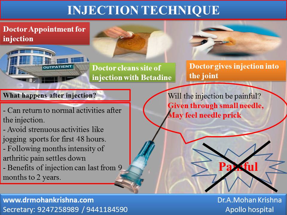
-          Steroid injections into the carpal may provide relief, but must be performed with utmost care. It is particularly important not to inject the median nerve itself any improvement may be temporary.
-          In persistent cases, surgery will be performed in order to reduce pressure on the large media nerve.

Surgery:

Surgery is usually advisable for persistent or worsening symptoms to prevent permanent loss of sensation and wasting of the muscles in the hand. In such cases without surgery, symptoms are likely to persist.
Surgical treatment usually involves dividing transverse carpal ligament in order to relieve the pressure on the median nerve. Freeing the nerve enables normal nerve conduction to resume. Traditionally, median nerve decompression was open surgical procedure, but also new technique of endoscopic carpal tunnel release is also being practiced.

Generally, both open and endoscopic techniques have excellent results although it may take few months for grip strength to return to normal.


Saturday, January 2, 2016

KNEE PAIN IN YOUNG ADULTS - CHONDROMALACIA PATELLA


CHONDROMALACIA PATELLA (ANTERIOR KNEE PAIN)

Introduction:

The most common location of knee pain in young adults is at the front of the knee joint. The problem usually arises from patella (Knee cap bone) and surrounding soft tissue.

Knee cap (Patella) is a small bone which lies in front of lower end of thigh bone (Femur). The back side of knee cap is lined by smooth tissue called cartilage. This covering helps the patella to glide in a groove on the lower end of thigh bone. The knee cap is held on the top by thigh muscles (Quadriceps) and lower part is connected to the shin bone by Patellar tendon. Function of knee cap is to aid knee movement and stabilize knee joint while walking.

Causes:

There are many theories suggesting softening and micro injuries to this lining resulting in pain. It could be due to repetitive micro trauma during falls, or vigorous unaccustomed exercises etc. Abnormal position of knee cap over the knee joint with or without weak ligaments may also damage the cartilage lining. As cartilage lining gets softened and roughened it may cause pain and crepitus (clicks and sounds) over the knee cap. Obesity makes the condition worse.

Complaints:

  • Pain in front of knee.
  • Pain on climbing up and down the steps or ramps.
  • Sudden ‘catch’ over the knee cap while getting up from the ground.
  • Dull aching pain after sitting for long time.
  • Discomfort during squatting (Using Indian toilet) and sitting cross legged.
  • Ocassional clicks and sounds over the knee cap.
  • Swelling.


Diagnosis:

Orthopaedician can make diagnosis based on your symptoms and examining the knee. Grating sensation behind knee cap might give us some idea of the roughness of patella when you tighten the knee cap muscles.
Some times there may be a little swelling the joint.

Tests:

Blood tests are normal.
X-rays are not helpful as the problem is with cartilage lining which cannot be seen on X-rays.
MRI (Magnetic resonance Imaging) scans can detect changes in the cartilage, but is used as last resort in unresponsive cases.
 Arthroscopy: It is possible to look inside the knee joint and back of the knee cap with pencil shaped camera (Arthroscope) to confirm the diagnosis. But arthroscopy is not done just for diagnosis and is only done if there is any need for surgery.

Treatment:

This condition generally improves over a period with some changes in the life style. But in some patients, symptoms persist for several years.
Analgesics (Pain Killers): Simple pain killers like Paracetomol and tramadol may be sufficient in most of the cases. Sometimes anti inflammatory medications (NSAIDS) may be necessary for a short period.
Nutritional supplements to cartilage lining like Glucosamine, Chondroitin, MSM and collagen peptides can be taken for 2 to 3 months which may relieve the symptoms.

Injections:

In some unresponsive cases Intra articualr Hyaluronic injections can help. They act by providing lubrication between joint surfaces and help to nourish cartilage lining of knee cap.

Physiotherapy:

Strengthening the anterior thigh muscles (Quadriceps) and muscles around the knee cap (VMO) helps to keep knee cap in right position.    

Surgery:

Surgical management is last resort in severe or unresponsive patients. It can be performed with arthroscope (Key hole surgery). The principle is to smoothen the rough surface of the back of knee cap. In some cases where there are large defects in the cartilage, multiple drill holes into the bone may help regrow some cartilage. Now a days we are combining this procedure with stem cell treatment in selective cases.

Do's & Dont's:

Please avoid any movement or position of which makes you feel the pain. It is better to avoid kneeling, squatting or sitting cross legged. One may go up and down the steps occasionally but avoid repeated stair climbing. Pain may get worse with exercise bicycle and walking on a treadmill with upward gradient (slope).

Sunday, November 22, 2015

HEEL AND FOOT PAIN

HEEL & FOOT PAIN

Heel pain is an extremely common complaint, and there are several common causes.
Plantar Fasciitis:  It is the most common condition that causes heel pain. Plantar fasciitis is due to irritation and inflammation of the tight tissue that forms the arch of the foot. The most common symptom is pain under the heel after a period of rest, i.e. severe pain and inability to walk in the morning after getting up from sleep. The same might happen after sitting for a long time and then trying to walk. Interestingly the pain gets better after a few steps though it may not go away completely.  Again, the pain may get worse with prolonged walking and standing. X ray may show a bony spur which may develop in long standing cases of heel pain due to plantar fasciitis, but it may not cause the symptoms.

Posterior Heel Pain:  Posterior heel pain causes symptoms behind the foot, rather than underneath. Posterior heel pain causes include Achilles tendinitis and retrocalcaneal bursitis. These patients may also develop bony growth behind the heel bone and when they are large may lead to rupture of Achilles tendon. Patients may have difficulty to walk comfortably on the affected side and pain that occurs at night or while resting.
Treatment Options:
Not all of these treatments are appropriate for every condition, but they may be helpful in your situation.
1.Rest - Avoiding the precipitating activity; for example, take a few day off jogging or prolonged standing/walking.  Rest can reduce the severity of pain and will allow the inflammation to begin to cool down.
2.Ice Packs - Icing will help to diminish some of the symptoms and control the heel pain. Icing is especially helpful after an acute exacerbation of symptoms.
3.Exercises and Stretches - Are designed to relax the tissues that surround the heel bone. Some simple exercises performed in the morning and evening, often help patients feel better quickly.
4.Anti-Inflammatory Medications - Help to both control heel pain and decrease inflammation.
5.Shoe Inserts - Are often the key to successful treatment of heel pain. The shoe inserts often permit patients to continue their routine activities without heel pain.
6.Injection therapy - In some cases, corticosteroid injections are used to help reduce the inflammation and relieve pain.
7.Removable walking cast - May be used to keep your foot immobile for a few weeks to allow it to rest and heal and is used only for severe cases.
8.Night splint - Wearing a night splint allows you to maintain an extended stretch of the plantar fascia while sleeping. This may help reduce the morning pain experienced by some patients.
9.Weight reduction – It helps to reduce stress on foot and heel and thus help to reduce the severity of pain.

If, after several months of non-surgical treatment, you continue to have heel pain, surgery will be considered.
Healthy Joint Club says:
Not being able to take first step out of bed in the morning can be frightening. More over severe pain from the heel may force you to restrict several day time activities. Except for a handful of cases and some uncommon conditions, most cases improve with treatment and time. Even though the time taken for the recovery may vary from person to person there is a natural tendency for these conditions to get better spontaneously.
STRETCHING EXERCISES
     
1.PLANTAR FASCIA STRETCH:
Sit in a chair and cross one foot over your other knee. Grab the base of your toes and pull them back toward your leg until you feel a comfortable stretch. Hold for 10 seconds and repeat 5 to 10 times.
2.ACHILLES STRETCH:
Stand on forefoot at the edge of stair, reach the step below with your heel, until you feel the stretch in the arch of the foot. Hold this position for 10 seconds and repeat 5 to 10 times.          
3.HEEL RAISE:
Stand behind the chair or any support and raise your body up onto your toes and hold for 5 seconds. Then slowly lower down yourself. Repeat 5 to 10 times.
4.TOWEL PICK:
Drop a towel on the ground and pick up the towel with your toes. Release and repeat 5 to 10 times.
5.FROZEN CAN ROLL:
Take a frozen juice can or tin, roll your feet on the can from front to back. Do it for 3 to 5 minutes.
6.TOWEL STRETCH:
Loop a towel around your toes and pull the foot towards your body keeping your leg straight. Hold this for 10 seconds and relax. Repeat 5 to 10 times.

Dr.A.Mohan Krishna
M.S.Ortho.,MCh orth (U.K)
Consultant Orthopedic surgeon,
Apollo Hospital
Appointments
Cell: 9247258989
         9441184590
email: bonesandjointsclinic@gmail.com
online appointments: 
www.drmohankrishna.com
www.bonesandjointsclinic.com


TAIL BONE PAIN - COCCYDYNIA


Pain in the tail bone can frustrate, irritate and embrace many patients for getting the treatment. This condition can be curable and prevented if proper care is taken.


Any irritation (inflammation) of the bony area (tail bone) situated between the folds of the buttocks is referred as Coccydynia.
The usual complaint is pain at the bottom region of the back on sitting. It is usually caused by injury, during pregnancy, chronic pressure on the tail bone due to sitting for long hours as of in those involved in driving occupation. Sometimes tail bone pain occurs in patients with chronic constipation. In one third of the cases the cause is not known.
Coccydynia is usually diagnosed based on the typical complaints and sometimes by an x-ray. Clinician can diagnose by examination and eliciting tenderness at the local region. It is necessary to directly visualize the tailbone region to rule out other causes and any infection. Sometimes infection of the hair follicles at the tailbone region can mimic coccydynia.
This condition usually frustrate patients and impair the quality of life.  Sometimes patients feel embarrassed to get the treatment. Treatment is usually by activity modification, anti inflammatory medications and local injections. Surgery is rarely indicated in non responsive cases.
Since long hours of sitting may aggravate the condition, a modified padded cushion with cut out at the back could relieve the pressure on the coccyx. (Doughnut cushion). Pelvic floor exercises could help to get relief from pain. Use of stool softeners and laxatives in cases of constipated patients could help to relive pain. Seitz bath by sitting in tub of hot water may provide pain relief. 

Oral anti inflammatory medications and local application of pain gels may tries to relive the symptoms. Local trigger injections with low dose steroid preparations will aid in complete relief in majority of cases. These local injections can be given by the doctor in the outpatient department.  In rare case surgical treatment by removal of coccyx may be required in those cases not responding to conservative methods.

Dr.A.Mohan Krishna
M.S.Ortho.,MCh orth (U.K)
Consultant Orthopedic surgeon,
Apollo Hospital
Appointments
Cell: 9247258989
         9441184590
email: bonesandjointsclinic@gmail.com
online appointments: 
www.drmohankrishna.com
www.bonesandjointsclinic.com